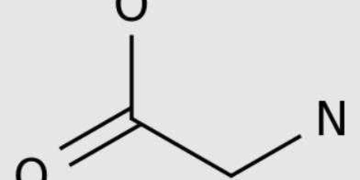
For nearly a century, researchers have known that cancer cells have a special appetite, consuming glucose in ways that normal cells do not. However, glucose uptake may only be part of the metabolic process of cancer. Researchers at the Broad Institute and Massachusetts General Hospital looked at 60 well studied cancer cell lines and analyzed which of the more than 200 metabolites were consumed or released by the fastest dividing cells. Their research produced the first large-scale cancer metabolic map and pointed out the key role of the smallest amino acid glycine in cancer cell proliferation. Their findings were published in the May 25th issue of the journal Science.
"There is increasing interest in the role of metabolism in cancer, but the research so far has focused on one or two very specific pathways," said senior author Vasisi Motta, co director of the Metabolism Program at the Broad Institute and a professor at Harvard Medical School and Massachusetts General Hospital. "We have taken an impartial approach to studying all metabolism, and the glycine pathway has emerged."
Mootha and his colleagues have developed a technique called CORE (consumption and release) analysis, which allows them to measure the flux of metabolites, precursors and products of chemical reactions occurring in the body. Most of the time, when researchers measure metabolites, they take a snapshot of their levels at a certain point in time. However, just as taking photos of highways cannot reveal the speed of traffic, such measurements cannot show which metabolites the cells are rapidly consuming or excreting.
"Using CORE, we can quantitatively determine how many metabolites each cell consumes or releases per hour," said co lead author Mohit Jain, a postdoctoral fellow at Mootha Lab. "We can now start calculating the flux or transport of nutrients into and out of cells."
The team applied CORE analysis to NCI-60, a collection of 60 cancer cell lines that have been studied by the scientific community for decades. These cell lines represent nine tumor types, and data on drug sensitivity, gene and protein activity, and cell division rate are publicly available. A summary of the team's information on metabolites has also been made public.
One of the most striking results of the new data is the relationship between glycine consumption patterns and the rate of cancer cell division. In the slowest dividing cells, a small amount of glycine is released into the culture medium. However, in rapidly dividing cancer cells, glycine is consumed in large quantities. The researchers pointed out that very few metabolites have this unusual pattern of "crossing the zero line", which means that rapidly dividing cancer cells consume metabolites, while slowly dividing cells actually release metabolites.
"We know very little about the metabolic activities that cause rapid or slow proliferation of cancer cells," Jain said. "But in these 60 cell lines, we clearly see a link between the speed of cell division and how much glycine they absorb."
"The CORE method is a screening exercise," said co lead author Roland Nilsson, who completed his postdoctoral work at the Mootha laboratory and is now at the Karolinska Institute. "This is a method for finding metabolic activity that may be interesting. You can use this data to conduct other experiments to verify it."
In addition to looking for metabolites related to the rate of cell division, the team also studied the expression of nearly 1500 metabolic enzymes. The enzymes required for the biosynthesis of glycine in mitochondria are most relevant.
"We have two independent methods - metabolite analysis and gene expression analysis - both of which indicate that glycine metabolism is important for proliferation rate," Mootha said.
To further validate and understand these results, the research team observed what happens when cancer cells are deprived of glycine, both by removing glycine from the culture medium and by blocking enzymes involved in glycine metabolism. In both cases, rapidly dividing cancer cells slow down, but slower growing cancer cells are not affected.
One limitation of observing this effect in laboratory cultured cancer cells is that these cells may behave differently in the human body. One way for researchers to track this work is to look at the research data of breast cancer patients in the past 25 years to find potential patterns between survival and levels of glycine metabolism related enzymes. They found that the higher the levels of these enzymes, the poorer the prognosis of patients.
The researchers envisioned many future directions for this work, including the wider application of CORE profiling.
Nielsen said: "This method provides a way to quickly understand specific cell types or tissues, so that you can see the conditions required for cell survival or growth." "We are interested in applying it to other environments, such as liver cells and muscle tissue, as well as diabetes and other diseases. There are many potential applications."


